
This comprehensive guide explores the anatomy, physiology, and mechanics of breathing, covering gas exchange, control systems, and common respiratory conditions․

Overview of the Respiratory System
The respiratory system’s primary function is facilitating gas exchange – taking in oxygen and expelling carbon dioxide․ This vital process supports cellular respiration and overall bodily function․ The system encompasses a network of organs, from the nasal passages to the alveoli within the lungs, working in concert to ensure efficient oxygen uptake․
Understanding its divisions – the conducting and respiratory zones – is crucial․ The conducting zone filters, warms, and humidifies air, while the respiratory zone is where gas exchange actually occurs․ Studying this system involves exploring anatomical structures, physiological mechanisms, and potential disruptions leading to respiratory diseases․ Resources like OpenStax and Osmosis provide detailed insights into these complex processes, aiding comprehension for students and healthcare professionals alike․
Functional Divisions: Conducting and Respiratory Zones
The respiratory system is functionally divided into two main zones: the conducting zone and the respiratory zone․ The conducting zone, including the nose, pharynx, larynx, trachea, bronchi, and bronchioles, serves to filter, warm, and humidify incoming air․ It doesn’t participate directly in gas exchange, but prepares the air for efficient uptake․
Conversely, the respiratory zone – comprised of the respiratory bronchioles, alveolar ducts, and alveoli – is where gas exchange between air and blood takes place․ This zone’s structure maximizes surface area for efficient oxygen absorption and carbon dioxide removal․ Understanding this division is key to grasping how the respiratory system effectively delivers oxygen to the body’s tissues, as highlighted in anatomy and physiology resources․
Organs of the Upper Respiratory System
The upper respiratory system initiates the breathing process, encompassing structures outside the chest cavity․ This includes the nose and nasal cavity, responsible for filtering, warming, and humidifying inhaled air․ The nasal cavity’s conchae increase surface area for these functions, while cilia and mucus trap particles․
The pharynx, or throat, serves as a passageway for both air and food, connecting the nasal cavity and mouth to the larynx and esophagus․ It’s divided into the nasopharynx, oropharynx, and laryngopharynx․ These organs work in concert to ensure clean, conditioned air reaches the lower respiratory system, preparing it for the vital exchange of oxygen and carbon dioxide, as detailed in anatomical studies․
Nose and Nasal Cavity
The nose serves as the primary entry point for air into the respiratory system, initiating the conditioning process․ Within the nasal cavity, conchae – superior, middle, and inferior – create turbulence, maximizing contact between air and the mucous membrane․ This membrane, rich in blood vessels, warms the incoming air․
Cilia, tiny hair-like structures, and mucus trap inhaled particles like dust and pollen, preventing them from reaching the lungs․ Olfactory receptors within the nasal cavity detect smells․ The nasal cavity is divided by the nasal septum, ensuring symmetrical airflow․ Proper function is crucial for efficient respiration and protecting the lower airways from irritants and pathogens․
Pharynx (Throat)
The pharynx, commonly known as the throat, is a crucial passageway shared by both the respiratory and digestive systems․ It’s divided into three regions: the nasopharynx, oropharynx, and laryngopharynx․ The nasopharynx lies posterior to the nasal cavity and contains the adenoids and auditory tubes․ The oropharynx, behind the oral cavity, handles both air and food․
Finally, the laryngopharynx connects to the larynx and esophagus․ During swallowing, muscles ensure the epiglottis covers the trachea, preventing food from entering the airways․ The pharynx plays a vital role in immune defense, housing tonsils that trap and neutralize pathogens․ Its strategic location necessitates coordinated function for both breathing and eating․
Organs of the Lower Respiratory System
The lower respiratory system begins with the larynx, or voice box, responsible for sound production via vocal cords․ Air then travels down the trachea, a rigid tube reinforced with C-shaped cartilage rings to prevent collapse․ The trachea bifurcates into the left and right primary bronchi, which enter the lungs․
Within the lungs, these bronchi branch repeatedly into smaller bronchioles, lacking cartilage but possessing smooth muscle allowing for bronchodilation and bronchoconstriction․ This branching forms the bronchial tree, ultimately leading to the respiratory zone where gas exchange occurs․ The lower system’s structures facilitate efficient air transport and regulation, crucial for oxygen uptake and carbon dioxide removal․
Larynx (Voice Box)
The larynx, commonly known as the voice box, is a cartilaginous structure situated between the pharynx and the trachea․ Its primary function is phonation, or sound production, achieved through the vibration of vocal cords․ These cords are bands of elastic tissue stretched across the laryngeal cavity․
The larynx also plays a vital role in airway protection, preventing food and liquids from entering the trachea during swallowing via the epiglottis․ Composed of several cartilages, including the thyroid cartilage (Adam’s apple) and cricoid cartilage, it provides structural support․ Adjusting vocal cord tension alters pitch, enabling diverse vocalizations․
Trachea (Windpipe)
The trachea, or windpipe, is a cartilaginous tube extending from the larynx to the bronchi, serving as the primary airway for air transport to and from the lungs․ Its structure consists of C-shaped rings of hyaline cartilage, providing support and preventing collapse during inhalation․ These rings are incomplete posteriorly, allowing for esophageal expansion during swallowing․
The tracheal lining is covered with a ciliated mucous membrane, which traps debris and pathogens, propelling them upwards towards the pharynx to be swallowed or expelled․ Smooth muscle within the trachea allows for adjustments in airway diameter․ This ensures efficient airflow and protects the lower respiratory system from irritants․
Bronchi and Bronchioles
The bronchi are formed by the division of the trachea into the right and left primary bronchi, each entering a lung․ These further divide into secondary and tertiary bronchi, progressively decreasing in diameter․ Their walls, like the trachea, contain cartilage for structural support, though the amount diminishes as they branch․
Bronchioles represent the smaller airways branching from the tertiary bronchi․ They lack cartilage but possess smooth muscle in their walls, allowing for bronchodilation and bronchoconstriction, regulating airflow․ Smooth muscle along the bronchi and initial bronchioles contains beta 2 adrenergic and muscarinic receptors, influencing airway diameter․ Ultimately, bronchioles lead to the respiratory bronchioles, initiating gas exchange․
Lungs: Anatomy and Function
The lungs are cone-shaped organs, occupying the thoracic cavity․ The right lung has three lobes, while the left has two, accommodating the heart․ Each lobe is further divided into lobules, separated by connective tissue․ Lungs are primarily composed of alveoli, facilitating efficient gas exchange․
Functionally, the lungs are responsible for oxygen intake and carbon dioxide expulsion․ This occurs within the respiratory zone, beginning with the respiratory bronchioles and culminating in the alveoli․ The lungs’ expansive surface area, created by millions of alveoli, maximizes gas exchange efficiency; Protective mechanisms, like mucus and cilia, defend against inhaled particles․
Alveoli and Gas Exchange
Alveoli are tiny, balloon-like structures within the lungs, representing the primary site of gas exchange․ Their thin walls, coupled with a dense capillary network, facilitate rapid diffusion of oxygen and carbon dioxide․ Type I alveolar cells form the structural component, while Type II cells secrete surfactant, reducing surface tension and preventing collapse․
Gas exchange occurs via simple diffusion, driven by partial pressure gradients․ Oxygen moves from the alveoli into the blood, binding to hemoglobin in red blood cells․ Simultaneously, carbon dioxide diffuses from the blood into the alveoli to be exhaled․ This vital process ensures oxygen delivery to tissues and carbon dioxide removal, maintaining homeostasis․
Muscles of Respiration
Effective ventilation relies on the coordinated action of several muscles․ The diaphragm, a primary muscle, contracts and flattens, increasing thoracic cavity volume during inhalation․ Relaxation allows it to return to its dome shape, decreasing volume during exhalation;
Intercostal muscles, located between the ribs, also play a crucial role․ External intercostals elevate the rib cage during inhalation, expanding the chest․ Internal intercostals depress the rib cage during forced exhalation․ Accessory muscles, like sternocleidomastoid and scalenes, assist during labored breathing, increasing thoracic volume․ Proper muscle function is essential for efficient gas exchange․
Diaphragm
The diaphragm is the principal muscle of respiration, a large, dome-shaped structure located at the base of the thoracic cavity․ During inhalation, it contracts, flattening and descending, which increases the volume of the chest cavity․ This creates negative pressure, drawing air into the lungs․
Conversely, during exhalation, the diaphragm relaxes, returning to its dome shape, reducing chest cavity volume and forcing air out․ Its innervation comes from the phrenic nerve, originating from cervical spinal nerves C3-C5․ Diaphragmatic weakness or paralysis significantly impairs breathing, necessitating mechanical ventilation in severe cases․
Intercostal Muscles
Intercostal muscles, located between the ribs, play a vital role in respiration, assisting the diaphragm in expanding and contracting the chest cavity․ There are three layers: external, internal, and innermost․ External intercostals elevate the rib cage during inhalation, increasing thoracic volume․
Internal intercostals depress the rib cage during forced exhalation, decreasing thoracic volume․ The innermost intercostals function similarly to the internal intercostals․ These muscles work synergistically with the diaphragm to facilitate efficient breathing․ Their coordinated action ensures adequate ventilation, delivering oxygen and removing carbon dioxide․
Pulmonary Ventilation: The Mechanics of Breathing
Pulmonary ventilation, commonly known as breathing, is the process of moving air into and out of the lungs․ It relies on pressure differences between the atmosphere and the alveoli․ Inhalation occurs when the diaphragm contracts and intercostal muscles expand the chest cavity, decreasing intrapulmonary pressure․ Air then flows into the lungs․
Exhalation is typically a passive process, driven by the elastic recoil of the lungs and relaxation of the diaphragm and intercostal muscles, increasing intrapulmonary pressure․ Forced exhalation involves contraction of abdominal and internal intercostal muscles․ Efficient ventilation is crucial for gas exchange and maintaining blood oxygen and carbon dioxide levels․

Gas Exchange: Oxygen and Carbon Dioxide Transport
Gas exchange is the vital process of transferring oxygen from the inhaled air to the blood, and carbon dioxide from the blood to the lungs․ This occurs in the alveoli, where oxygen diffuses across the thin alveolar and capillary walls into the bloodstream․ Simultaneously, carbon dioxide moves in the opposite direction․
Oxygen transport primarily relies on hemoglobin within red blood cells․ Carbon dioxide is transported in three ways: dissolved in plasma, bound to hemoglobin, and as bicarbonate ions․ Efficient gas transport ensures tissues receive adequate oxygen and waste carbon dioxide is removed, maintaining cellular function and overall homeostasis․
Control of Breathing
Breathing control is a complex process regulated by the brainstem, specifically the medulla oblongata and pons․ The medulla sets the basic rhythm of breathing, while the pons fine-tunes it․ Chemoreceptors play a crucial role, monitoring blood levels of oxygen, carbon dioxide, and pH․
Central chemoreceptors in the brainstem respond to changes in carbon dioxide and pH, while peripheral chemoreceptors in the carotid and aortic bodies detect oxygen levels․ These receptors send signals to the brainstem, adjusting breathing rate and depth to maintain optimal blood gas levels and pH balance, ensuring adequate oxygen delivery and carbon dioxide removal․
Role of the Brainstem
The brainstem, comprising the medulla oblongata and pons, is central to breathing control․ The medulla contains the respiratory center, establishing the basic rhythm of inhalation and exhalation․ This center generates nerve impulses that travel to respiratory muscles, initiating contractions․
The pons modifies the medullary rhythm, smoothing transitions between inspiration and expiration․ It also influences the depth and rate of breathing․ Damage to the brainstem can severely impair or halt breathing․ Furthermore, the brainstem integrates input from chemoreceptors and other sensory receptors, adjusting ventilation to meet the body’s metabolic demands, ensuring proper oxygen and carbon dioxide levels․
Chemoreceptors and Regulation
Chemoreceptors play a vital role in regulating breathing by monitoring blood gas levels․ Central chemoreceptors, located in the medulla oblongata, primarily detect changes in pH and carbon dioxide (CO2) levels in the cerebrospinal fluid․ Increased CO2 leads to a decrease in pH, stimulating increased ventilation․
Peripheral chemoreceptors, found in the carotid and aortic bodies, sense changes in oxygen (O2), CO2, and pH in the blood․ A significant drop in O2, or increases in CO2 or decreased pH, trigger signals to the brainstem, prompting adjustments to breathing rate and depth․ This feedback loop maintains blood gas homeostasis, ensuring adequate oxygen delivery and carbon dioxide removal․
Respiratory Volumes and Capacities
Respiratory volumes describe the amount of air moved during different phases of breathing․ Tidal volume (TV) is the air inhaled or exhaled during normal breathing, typically around 500ml․ Inspiratory reserve volume (IRV) is the extra air inhaled forcefully, while expiratory reserve volume (ERV) is the extra air exhaled forcefully․
Respiratory capacities combine two or more volumes․ Vital capacity (VC) is the total air exhaled after maximal inhalation (TV + IRV + ERV)․ Functional residual capacity (FRC) is the air remaining in the lungs after a normal exhalation (ERV + residual volume)․ Total lung capacity (TLC) represents the total air the lungs can hold (VC + FRC)․
Common Respiratory Diseases and Disorders
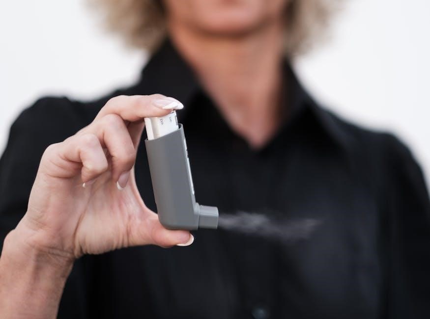
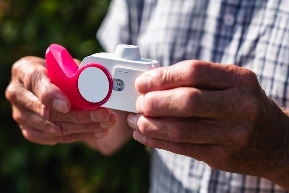
Asthma is a chronic inflammatory disease of the airways, causing recurring episodes of wheezing, chest tightness, shortness of breath, and coughing․ Bronchoconstriction, inflammation, and mucus production characterize it, often triggered by allergens or irritants․
Chronic Obstructive Pulmonary Disease (COPD) encompasses conditions like emphysema and chronic bronchitis, typically caused by long-term exposure to irritants, most commonly cigarette smoke․ It’s marked by airflow limitation and difficulty breathing․ Both diseases significantly impair lung function, reducing quality of life and potentially leading to respiratory failure․ Early diagnosis and management are crucial for both conditions․
Asthma
Asthma is a chronic inflammatory disease affecting the airways, causing recurring episodes of wheezing, shortness of breath, chest tightness, and coughing․ This inflammation leads to airway hyperresponsiveness, meaning the airways narrow easily in response to various triggers like allergens, exercise, or cold air․
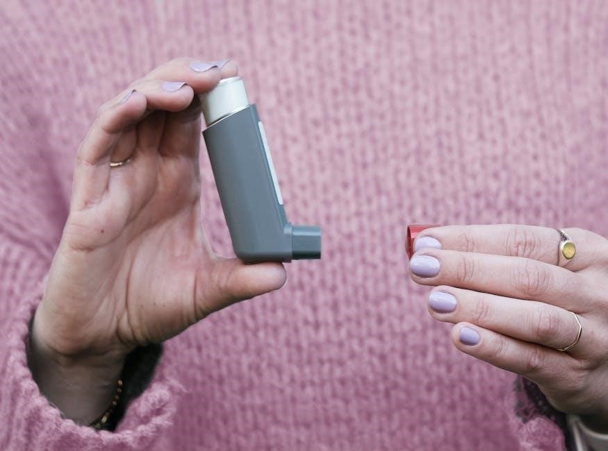
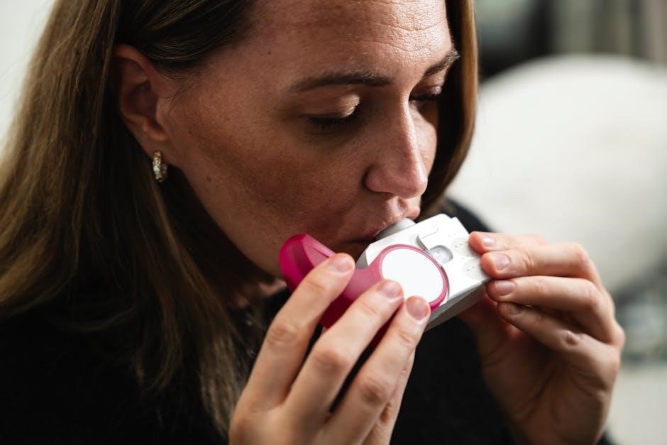
Pathophysiology involves bronchoconstriction, airway edema, and increased mucus production․ Diagnosis typically includes a physical exam, pulmonary function tests (spirometry), and allergy testing․ Treatment focuses on both long-term control (inhaled corticosteroids) and quick relief (bronchodilators like beta-2 agonists) to manage symptoms and prevent exacerbations․ Proper management is vital for maintaining a normal lifestyle․
Chronic Obstructive Pulmonary Disease (COPD)
COPD encompasses progressive lung diseases, most commonly chronic bronchitis and emphysema, that block airflow and make breathing difficult․ Chronic bronchitis involves long-term inflammation and mucus production in the airways, while emphysema damages the alveoli, reducing gas exchange efficiency․
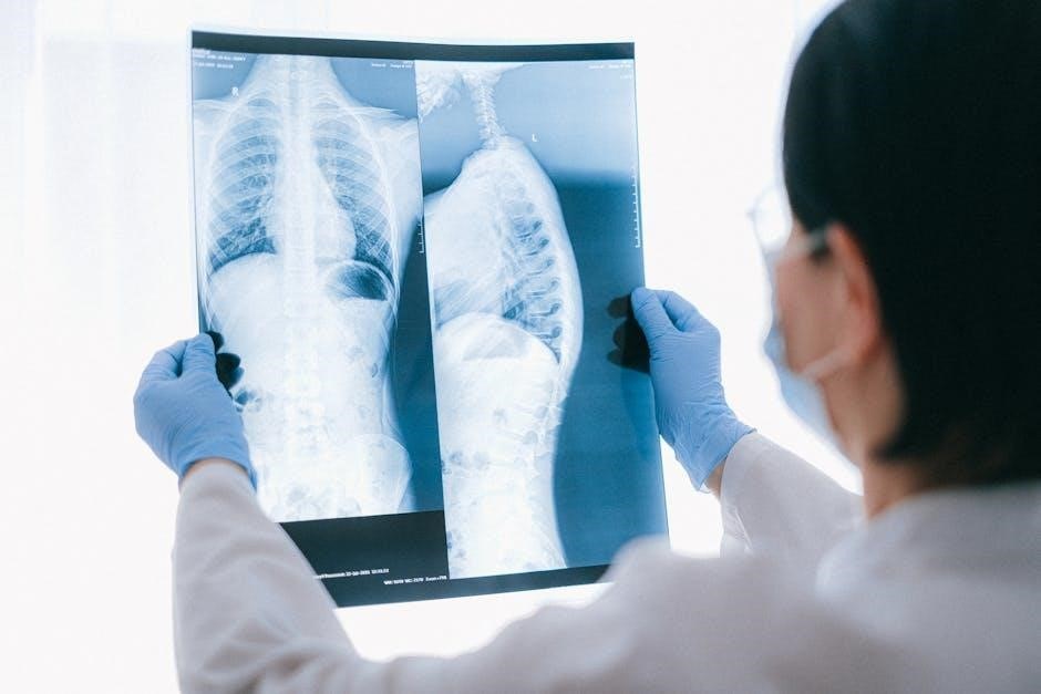
Smoking is the primary cause, though long-term exposure to irritants like air pollution can also contribute․ Symptoms include chronic cough, excessive mucus, wheezing, and shortness of breath․ Diagnosis relies on spirometry and imaging․ Treatment focuses on symptom management with bronchodilators, inhaled corticosteroids, and pulmonary rehabilitation; oxygen therapy may be needed in severe cases․
Receptors in the Respiratory System
The respiratory system utilizes various receptors to regulate airflow and respond to changes in blood gases․ Beta 2 adrenergic receptors, found in the smooth muscle of the trachea and bronchi, cause bronchodilation when stimulated by epinephrine, opening airways․ Conversely, muscarinic receptors mediate bronchoconstriction when activated by acetylcholine, narrowing airways․
These receptors are crucial targets for medications․ Beta 2 agonists, like albuterol, relax airway muscles, relieving asthma symptoms․ Anticholinergics block muscarinic receptors, also promoting bronchodilation․ Understanding these receptor interactions is vital for managing respiratory conditions and optimizing treatment strategies, ensuring effective airflow regulation․

Beta 2 Adrenergic Receptors
Beta 2 adrenergic receptors are prominently located in the smooth muscle surrounding the trachea and the initial branches of the bronchi․ These receptors play a critical role in regulating airway diameter, responding to stimulation by catecholamines like epinephrine․ When activated, they initiate a signaling cascade leading to smooth muscle relaxation and subsequent bronchodilation – widening of the airways․
This mechanism is fundamental in responding to physiological stressors and is a key target for pharmacological intervention․ Beta 2 agonists, commonly used in asthma treatment, mimic epinephrine’s effect, providing relief by easing airflow obstruction․ Understanding their function is crucial for comprehending respiratory control and therapeutic strategies․
Muscarinic Receptors
Muscarinic receptors, also present in the smooth muscle of the trachea and early bronchi, contrast with beta 2 adrenergic receptors in their function․ These receptors are activated by acetylcholine, a neurotransmitter released by the parasympathetic nervous system․ Upon activation, muscarinic receptors trigger smooth muscle contraction, leading to bronchoconstriction – narrowing of the airways․
This response is part of the body’s “rest and digest” state, but can be problematic in respiratory conditions․ Anticholinergic medications, which block muscarinic receptors, are sometimes used to counteract bronchoconstriction and improve airflow․ Understanding the opposing roles of beta 2 and muscarinic receptors is vital for grasping the complex regulation of airway diameter and effective treatment strategies․
Animated Videos and Quizzes for Learning
Enhance your understanding of the respiratory system with a wealth of interactive resources! Numerous animated videos visually demonstrate complex processes like pulmonary ventilation, gas exchange within alveoli, and the mechanics of breathing․ These dynamic tools clarify difficult concepts, making learning more engaging and effective․
Complementing the videos are practice quizzes, readily available online․ Georgia Highlands College’s LibGuides, for example, offers a chapter twenty-two practice quiz․ These assessments allow you to test your knowledge, identify areas needing further study, and reinforce key concepts․ Osmosis also provides comprehensive notes and diagrams․ Utilize these resources to solidify your grasp of respiratory anatomy and physiology, preparing you for success!